Isolation and culture of ADSCs
The lipoaspiration technique was used to obtain adipose tissue samples from healthy subjects after their written informed consent. The samples were washed with warm phosphate-buffered saline (PBS) to eliminate blood and oil. Next, the adipose tissue was digested using 0.025% type I collagenase (Worthington Biochemical Corporation, Lakewood, NJ, USA) for 1 h at 37 °C. The lipoaspirate was centrifuged at 2000 rpm for 5 min to separate the pellet containing ADSCs, and the pellet was resuspended in stromal medium (DMEM-LG; Gibco, Carlsbad, CA, USA) supplemented with 10% fetal bovine serum (Sigma-Aldrich, St. Louis, MO, USA), 1% Penicillin–Streptomycin (Gibco, Carlsbad, CA, USA), and 1% Glutamax (Gibco, Carlsbad, CA, USA). The cells were seeded at a density of 103 cells/cm2 and then incubated at 37 °C and 5% CO2. After 48 h, the nonadherent cells were removed, and the remaining cells were maintained until they reached 80%–90% confluence, while the medium was changed every three days. ADSCs were harvested using 0.25% trypsin–EDTA (Gibco, Carlsbad, CA, USA), and the cultures were either expanded or cryopreserved for future use.
Surface modification and cell culture
To determine the effects of adhesion molecule as coating materials for ADSCs culture in vitro, the experiments were divided into three groups, ADSCs were cultured on uncoated surface as the control group (CTRL), coated surface with 5 µg/cm2 FN (Merck, Darmstadt, Germany) and 0.5 µg/cm2 VN (Advanced BioMatrix, Carlsbad, CA, USA). In the FN-coated group, 1 mL FN was added to a 35 mm dish, followed by the removal of excess solution. Air dried for at least 45 min at room temperature (RT) was performed. In the VN-coated group, 1 ml VN was added to 35 mm-dish, incubated for 1 h at RT, and then washed with PBS twice. To investigate the cellular senescence, 1 × 10⁵ ADSCs at the 2nd passage (P2) were seeded on uncoated (CTRL) and coated surface. ADSCs were cultured at 37 °C in 5% CO2 until the cells reached the 5th passage (P5), 7th passage (P7), and 10th passage (P10). The biological activities of ADSCs under each condition were evaluated at P5, P7, and P10. The MDM2 inhibitor, Nutlin-3a (Sigma-Aldrich, Taufkirchen, Germany), was dissolved in DMSO (PanReac Applichem, Darmstadt, Spain). Then 10 µM Nutlin-3a was added to the ADSCs culture at P10. This was followed by incubation for 48 h before concluding the experiment.
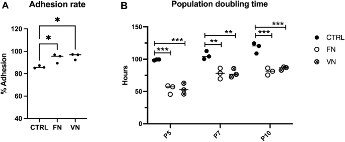
Cell adhesion assay
1 × 10⁵ cells were seeded in a 24-well plate coated with FN and VN to evaluate the adhesion properties of ADSCs. Here, ADSCs cultured on an uncoated surface were used as control. The seeded cells were incubated for 12 h in a humidified atmosphere containing 5% CO2 at 37 °C, and then the nonadherent cells were carefully removed and discarded. The adherent cells were detached using 0.25% trypsin–EDTA (Sigma-Aldrich, St. Louis, MO, USA) and manually counted using a hemocytometer. The percentage of cell adhesion was calculated using the following equation to assess cell adhesion rate:
$${text{Cell adhesion rate }}left( % right) , = , left( {{text{Number of adherent cells}}/{text{Number of seeding cells}}} right) times {1}00.$$
Proliferative activity
ADSCs were seeded on control, FN, and VN-coated dishes at a density of 1 × 10⁵ cells to investigate the impact of adhesion molecule-coated surfaces on cell growth. Upon ADSCs cultured reaching 90% confluence, the cells were detached by trypsinization, and their viability was assessed using trypan blue staining. The proliferative activity of ADSCs was assessed by calculating the population doubling time (PDT) using the following expression: PDT = CT/PDN, where CT denotes the culture time in hours. PDN denotes the number of population doublings and is calculated using the formula: PDN = [logNH − logNI]/log2, where NI and NH represent the initial cell seeding number and the number of harvested cells, respectively. The PDT of ADSCs cultured on an uncoated surface was used as the control group for comparison.
Senescence-associated β-galactosidase (SA-β-gal) staining
SA-β-gal activity was assessed using a senescence cell staining kit (Cell Signaling Technology, Danvers, MA, USA) to investigate the impact of coated surfaces of adhesion molecules on cellular senescence. Specifically, 3 × 10⁴ ADSCs at P5, P7, and P10 were seeded in 35-mm culture dishes with coated surfaces using FN and VN. Then, the cells were cultured in a humidified atmosphere containing 5% CO2 at 37 °C until the cultures reached 60% confluence. The cells were washed with PBS and then fixed for 15 min at RT before being stained with SA-β-gal staining solution in a dry incubator at 37 °C for 16–18 h. The formation of a blue color, which represented senescent-positive cells, was observed using an inverted microscope (Olympus, Shinjuku, Tokyo, Japan). The number of SA-β-gal-positive cells was counted and presented as the percentage of SA-β-gal activity using the following formula: SA-β-gal activity (%) = (Number of SA-β-gal-positive cells/Number of total cells) × 100.
RNA extraction and quantitative real-time PCR
RNA was extracted using the phenol–chloroform procedure from the P5, P7, and P10 of ADSCs grown under individual conditions in TRIzol reagent (Invitrogen, Waltham, MA, USA). The RNA concentration was measured using a nanodrop spectrometer (Thermo Scientific, Waltham, MA, USA). cDNA was synthesized from 1 µg of RNA using iScript reverse transcription (Bio-Rad Laboratories, CA, USA), and the mRNA levels of p16, p21, p53, ITGA5, ITGAV, ITGB1, ITGB3 and ITGB5 were examined using quantitative real-time PCR. Each cDNA sample was mixed with a PCR master mix containing target gene forward and reverse primers (Table 1), nuclease-free water, and SYBR FAST qPCR master mix (KAPA Biosystem, Wilmington, MA, USA). Glyceraldehyde-phosphate dehydrogenase (GAPDH) was used as an internal reference control to normalize the expression levels of the genes of interest. Finally, the difference in transcript levels of senescence-associated genes was calculated using the comparative CT method (2−∆∆Ct) and presented as a relative gene expression to the control group.
Western blotting analysis
For immunoblotting, the protein from ADSCs culture in different coated surfaces was dissolved in lysis buffer (Merck, Darmstadt, Germany) with protease inhibitors (Merck, Darmstadt, Germany), and the concentration was determined by Bicinchoninic acid (BCA) Protein Assay Kit (Thermo Scientific, Waltham, MA, USA). The 20 µg of protein samples were separated on a 10% polyacrylamide gel and transferred to an Immobilon-P transfer membrane (Merck Millipore, Burlington, MA, USA) using the Mini-Protean© system (Bio-Rad Laboratories, Feldkirchen, Germany). The membranes were blocked in 5% skimmed milk (Merck, Darmstadt, Germany) for 2 h and incubated with the primary antibodies at 4 °C overnight. The primary antibodies used were AKT (#4691S), MDM2 (#86934S), p21 (#2947S), p53 (#2524S) (Cell Signaling Technology, Danvers, MA, USA), and β-Actin (Merck, Darmstadt, Germany) as loading controls for normalization. In this process, the membranes were cut before hybridization with various antibodies, resulting in the absence of images with adequate length.
On the following day, the membranes were incubated using an ECL Prime Western Blotting Detection Reagent (GE Healthcare, Buckinghamshire, UK) at RT for 2 h with secondary antibodies including anti-mouse IgG-HRP (#7074P2) and anti-rabbit IgG-HRP (#7076S) (Cell Signaling Technology, Danvers, MA, USA). The signal was then detected and analyzed via the ChemiDoc™ MP Imaging System (Bio-Rad Laboratories, Los Angeles, CA, USA).
Immunofluorescent study
ADSCs samples were fixed in warm 4% paraformaldehyde for 15 min at RT and then washed three times with PBS. The samples were then permeabilized with 0.025% Triton X-100 (Sigma-Aldrich, St. Louis, MO, USA) for 10 min and blocked in 5% bovine serum albumin (BSA) (Sigma-Aldrich, St. Louis, MO, USA) in PBS for 1 h at RT. The samples were incubated overnight at 4 °C with primary antibodies, i.e., rabbit anti-HMGB1 (#3935S, 1:200) and rabbit IL-6 (#12912S, 1:200) (Cell Signaling Technology, Danvers, MA, USA), diluted in 1% BSA in PBS. The samples were then incubated with secondary antibody solutions, i.e., Alexa Fluor 488 goat antirabbit (AB150077, 1:250) (Abcam, Cambridge, UK), for 45 min at RT. Finally, the samples were counterstained with Prolong Gold™ Antifade Reagent with DAPI (Thermo Scientific, Waltham, MA, USA) for 24 h at RT in the dark. The samples were photographed using a fluorescence microscope (Olympus BX51, Shinjuku, Tokyo, Japan).
Statistical analysis
The data are presented as mean ± standard deviation (SD) of at least three individual experiments (the Supplementary Figs. S1, and S3 are presented from two individual experiments). Statistical analysis was performed using an ordinary one-way ANOVA of GraphPad Prism 9.0 software (GraphPad Software, San Diego, CA, USA). A p-value < 0.05 was considered statistically significant.
Statement
Human adipose tissue samples from healthy subject was approved by the Mahidol University Central Institutional Review Board and was performed in accordance with the Declaration of Helsinki (MU-CIRB 2018/202.1441) with the title “Study of Mesenchymal Stem Cells Senescence Derived Adipose Tissue”. There are no clinical trials or animal experiments in our research. All experiments were performed in accordance with relevant guidelines and regulations.
- SEO Powered Content & PR Distribution. Get Amplified Today.
- PlatoData.Network Vertical Generative Ai. Empower Yourself. Access Here.
- PlatoAiStream. Web3 Intelligence. Knowledge Amplified. Access Here.
- PlatoESG. Carbon, CleanTech, Energy, Environment, Solar, Waste Management. Access Here.
- PlatoHealth. Biotech and Clinical Trials Intelligence. Access Here.
- Source: https://www.nature.com/articles/s41598-024-65339-z